| 
Electroimpedance diagnosis of the breasts cancer
The new medical device electrical impedance mammography "MEIK" is
concentrate mainly on the early discover of pathology of the milk
gland, also the breasts. The oncological pathologies of the breasts
are permanent in the center of attention as of the world health society.
The reason is the ceaseless growth of oncological diseases of breasts
and from that the following also a high mortality of female patients
with the carcinoma pathology of the breast. During the last 15 years
has the carcinoma of the breast moved from the fourth place to the
first place in the structure of oncological pathology. Each fifth
woman is deceasing on the carcinoma of the breast. The success of
the actual treatment nearby clear depends from the stadium of the
development of the discovered cancer process. So as early as possible
identification of the carcinoma or also other diseases of the breast
can lead to a significant reduction of proportion of mortality between
the woman.
At the present the most used method of diagnosis by the breasts is
the radiological rtg mammography . The actual examination and also
the method are connected with many complications, trouble and accessory
effects for the female patient. It´s mainly about the actual process
of examination, where the breasts are pressed between two metal plates,
what is painful enough. Successive the breast will be treated with
rtg radiation. But many scientist experts consider also small doses
of rtg radiation for the initiator of cancer growth. The classic rtg
mammography is nearby very little effective by younger female patients
(under 40 years). The reason is the higher density of the soft tissue
in the breast.
Research in the area of technology of electric impedance opened new
perspectives in research of electrical conductivity of human tissue
and with it also of the milk gland. The electric resistance of the
bio tissue (conductivity) nearby belongs between it´s main characteristics.
The last scientific research in the area of conductivity utilised
the electroimpedance of alive tissues for the construction of the
electrical impedance tomography (EIT) and with it also the mammograph,
which is able to visualize the soft tissues in the human organism.
Changes of the physiological status of organs under the influence
of chemical and physical factors as also various pathological processes
have a clear influence to the electrical properties of the alive tissue.
The electroimpedance tomograph (mamograph) is about all able to visualize
the physiological status of the tissue, then also organs, which are
defined through biological processes in the organism and mainly these,
which are manifested through the change of the electrical conductivity,
as are for example tumor processes.
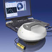
The output from this device is a graphic and digital information (electrical
conductivity, impedance and histogram) about the status of organs
and tissues.
The single examination is absolute secure for patient and also for
the operating staff. The examination can be repeated many times in
succession and that is very suitable by judgement of the influence
of the single treatment on the status of the pathological process
mainly by oncological patients.
Electroimpedance diagnostic method doesn´t show any accessory influences
on the alive organism. This method is absolute secure.
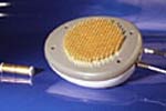
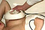
The single measurement of the electroimpedance will be carried out
with the apposing of the measure probe of the device to the breast
so, that as much as possible the number of small electrodes are in
contact with the skin.
Nearby one measurement lasts approximately 20 seconds and the signal
will be successive elaborated and visualized. The computer elaboration
of the signal clearly ease doctors to distinguish the single pathologies
and following to determinate the diagnosis by the examined female
patient.
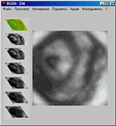
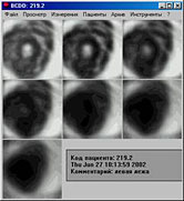 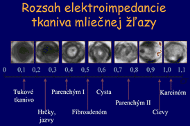
The chart output from the electroimpedance mammograph make it possible
to observe the status of the measured tissue of some seven transversal
cuts deep until 4,5 cm and observe the output parameters separate
for each stratum.
Through the combination of examinations with help of electroimpedance
mammograph, microwave radiometry and sonography it´s possible to reach
a maximal reliability exactitude, specificity as also sensibility
of the diagnostic method of the carcinoma of milk gland (breast),
but also other organs of the human body. |